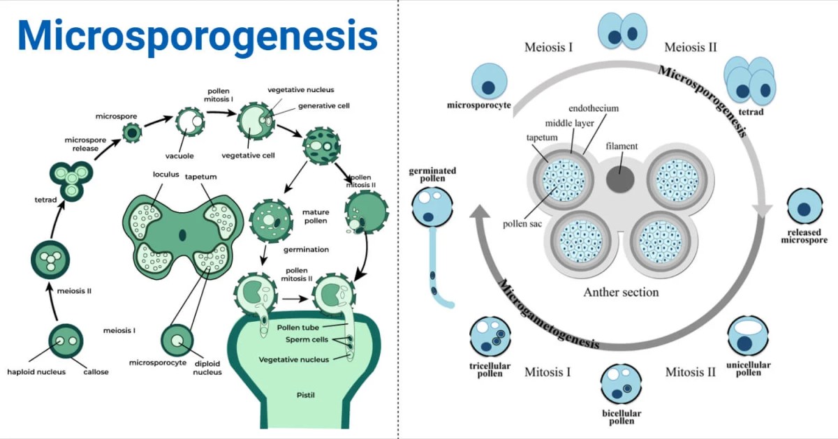
The process of formation of microspores or pollen grains inside the pollen sac or microsporangium is called microsporogenesis.
Interesting Science Videos
Significance of Microsporogenesis
Maintains ploidy– The process of microsporogenesis produces four haploid microspores from the diploid microspore mother cells. This maintains the chromosome number in the offspring and the diploid number when fused with the egg.
Causes genetic variation– The meiotic process of microsporogenesis enables the random segregation and crossing over of chromosomes. This creates differences in the genetic makeup of the male gametes compared to the parent plant.
Male fertility– Microsporogenesis is responsible for male fertility and necessitates the formation of both somatic and reproductive cells in the anther.


Structure of Stamen
A stamen in a flower consists of two parts, an anther, and a filament.
The anther is the bulbous top of the stamen that produces and stores pollen grain. It is generally bilobed or bithecous, connected by a strip of tissue known as connective. These two lobes consist of four sac-like structures known as microsporangia.
In some plants belonging to the Malvaceae family, anther is monoecious with two microsporangia.
The filament is the stalk-like structure that supports the anther. The proximal end of the filament is attached to the thalamus.
T.S. of anther shows parietal anther walls and microsporangia. The anther wall is made up of a single-layered epidermis and is protective in function.
Structure of microsporangium
The microsporangium lies inside the anther wall and is divided into three layers-
Endothecium
This is a single layer that lies just beneath the epidermis and has different walls. The outermost walls are thin but the inner walls possess radial thickenings due to the presence of cellulose fibers. Callose bands are also present in the inner walls, however, at some points, these bands are absent which is known as stomium which exactly lies at the junction of two pollen sacs. The dehiscence of anther takes place from this place. The endothecium layer is hygroscopic due to the presence of fibrous thickening which helps in pollen dehiscence.
Middle layer
This layer is a 1-3 thick-celled structure and is made up of parenchymatous cells. However, the middle layer is ephemeral in nature and degenerates during maturity. Food is primarily stored in this layer by parenchymatous cells.
Tapetum
It is the innermost and single layer of the cell that contains dense cytoplasm and a prominent nucleus. It absorbs food from the middle layer and provides nutrition to the microspores. This layer secretes hormones and enzymes and is usually diploid but due to endomitosis, they become polyploid.
It is of two types-
- Ameboid type and
- Glandular type
Ameboid type– It is also known as periplasmodial tapetum and is mainly found in primitive angiosperms. In this type, the cells undergo repeated karyokinesis without the division of cytoplasm resulting in a multinucleate mass of cytoplasm called plasmodium.
Glandular/ secondary tapetum- It is the developed type of tapetum and does not degenerate quickly, rather it secretes enzymes and hormonal substances. They form granular bodies before degeneration and are known as Pro-ubisch bodies or orbicules.
Inside these layers lie the pollen tetrads.
Process of Microsporogenesis
The young anther consists of a mass of meristematic cells beneath the epidermis. The cells lying at the four corners become enlarged, radially elongated, and prominent and are now known as archesporial cells. Simultaneously, vascular tissues are also formed in the middle portion of the anther.
The archesporial cell undergoes periclinal division to form two layered cells- the outer one towards the epidermis is known as primary parietal cells and the inner one towards the inner side is called primary sporogenous cells.
The primary parietal cells form the secondary parietal layers which undergo both anticlinal and periclinal divisions to form endothecium, middle layer, and tapetum.
According to Davis 1966, there are various ways of anther wall development. They are as follows:
Basic type– It is the most primitive type of anther wall development in which there are two layers of secondary parietal cells. The outer layer forms endothecium and one middle layer and the inner layer forms one middle layer and tapetum.
Dicotyledonous type– In this type, the outer secondary parietal layer forms endothecium and one middle layer, and the tapetum is formed by inner secondary parietal layers.
Monocotyledonous type– In this type, endothecium is formed from outer secondary parietal layers and one middle layer and tapetum is formed by inner secondary parietal layers.
Reduced type- In this type, the middle layer is absent. The outer secondary parietal layer gives rise to endothecium and the inner layer gives rise to tapetum.
The primary sporogenous cells divide mitotically to form sporogenous cells and later it is differentiated into microspore mother cells (MMC) or pollen mother cells (PMC) during the formation of anther wall and pollen sac.
The pollen mother cell undergoes meiosis to form 4 haploid microspores which are attached by a callose wall and are known as microspore tetrad.
With the entry of the microspore mother cell into meiosis, the wall of the PMC gets thickened due to the deposition of callose.
There are two types of microsporogenesis based on cytokinesis during meiosis-
Successive type– The two nuclear divisions or karyokinesis are followed by successive cytokinesis. The wall formation takes place in the center and extends towards the periphery. The tetrads formed by successive wall formation are called isobilateral tetrads and are common in monocots.
Simultaneous type– In this type, the wall is formed only after the completion of karyokinesis in both meiosis I and II. The wall formation occurs from the periphery towards the center. The tetrad formed by simultaneous wall formation is called tetrahedral tetrad and is common in dicots.
Usually, the microspore tetrads are separated shortly after meiosis. The tapetum secretes the callase enzyme that leads to the breakdown of the callose wall, thus releasing the microspores.
Types of Microspore Tetrad
Isobilateral– It is a common type of pollen tetrad in monocots where the four microspores are arranged at the four corners of the square in one plane. Example- Zea mays
Tetrahedral– In this type, the microspores are arranged in a tetrahedral manner and are mostly found in dicots. Example- Rhododendron.
Decussate– In this type, four pollens are arranged in two groups that lie perpendicular to each other. Example-Magnolia
Linear– It is a type of tetrad in which four pollen lie in a straight line. Example- Mimosa pudica
T-shaped– Here, the two microspores are arranged in a transverse plane and two in a longitudinal plane. Example- Aristolochia elegans
Aristolochia elegans has all 5 types of tetrads.
Microspore tetrad morphology can help determine the phylogeny of plant species.
Compound pollen grains–
In some cases, the microspore tetrad is unable to separate into single-celled pollen grains and thus remain together. These are called compound pollen grains. Examples are Annona, Typha, Drosera, etc.
Pollinia-
In families like Orchidaceae and Asclepiadaceae, the pollens of each anther lobe cohere in a single mass called pollinia or massula. Each pollinium has a stalk known as a Caudicle and a sticky base or disc known as a corpusculum. The caudicle and corpusculum together are termed a translator apparatus.
Polyspory-
The formation of more than four microspores is called polyspory.
For example in Cuscuta reflexa, the presence of 11 microspores has been reported.
Factors Influencing Microsporogenesis
The major factors influencing microsporogenesis are-
Environmental factors–
Environmental factors are one of the determinant factors in controlling microsporogenesis and stressful conditions can disrupt normal pollen development. These are as follows-
Temperature– High temperatures can disrupt meiosis and also may lead to defective chromosomal division, microspore degradation, and pollen sterility. Many crops such as wheat, and rice have reduced pollen viability due to heat stress. Cold stress can disrupt spindle fiber formation and cause incomplete separation of chromosomes.
Humidity- Adequate humidity is required for anther dehiscence and pollen maturation. Too low humidity can lead to premature dying of the anther and microspores, while high humidity can encourage microbial infection.
Water stress– Drought conditions may limit nutrient and water supply impairing cell division and differentiation. Flooding may inhibit cellular respiration thus affecting pollen quality.
Light intensity and photoperiod– Light intensity and photoperiod are very crucial for pollen development. It affects the availability of resources and an aberrant photoperiod may lead to delay in microsporogenesis.
Pollution– Exposure to pollutants such as sulfur dioxide, and heavy metals may cause damage in anther development, and may decrease pollen viability.
Genetic factors–
It is a crucial means to ensure the production of healthy microspores. The genes control each of the steps involved in the process of pollen development. Mutation or abnormal expression of such genes causes abnormalities.
For example, mutations in TDR (Tapetum Determinant 1) affect tapetum function and result in pollen sterility.
Chromosomal aberrations– Aneuploidy may occur due to non-disjunction, inversion, or translocation in meiosis. Polyploidy may lead to chromosome incompatibility.
Genetic Sterility– Some plants show natural male sterility caused by nuclear or cytoplasmic genes. This type of sterility leads to the failure of viable pollen to fertilize ovules.
Physiological Factors– Physiological factors including hormonal balance, metabolic activities, and energy supply, play a significant role in the regulation of microsporogenesis.
Plant hormones– Hormones like Gibberellin help in the growth of stamens and anthers. Abscisic acid regulates the tolerance of drying in developing pollen grains. An imbalance in these hormones may disrupt meiosis and pollen maturation.
Tapetal function– Abnormal tapetal development or premature degeneration can lead to pollen abortion.
Biotic factors– Biotic factors such as fungal, bacterial, and viral pathogens and pests may interfere with the development of microspores thus leading to infections. Insects feeding upon floral tissues can physically interfere with anther or reduce pollen production.
Tapetum Role in Microsporogenesis
The tapetum is the innermost layer of the anther wall. It is involved in microsporogenesis. It supplies nourishment to the developing microspores and secretes enzymes that help break down the callose wall. Tapetum also participates in the formation of the pollen wall by providing sporopollenin precursors. These make the exine highly resistant to environmental stress. The tapetal cells are metabolically active and are important for the development of viable pollen grains.
Comparison between microsporogenesis and megasporogenesis
- Microsporogenesis is the formation of microspores in the anther whereas megasporogenesis is the formation of megaspores in the ovary.
- The arrangement of microspore tetrad is usually tetrahedral whereas megaspore tetrad is commonly linear.
- All four microspores of microspore tetrad are functional whereas only one megaspore of a spore tetrad is functional.
- During microsporogenesis, all the sporogenous cells differentiate into microspore mother cells whereas during megasporogenesis, only one sporogenous cell differentiates into a megaspore mother cell.
Microgametogenesis
Microgametogenesis is defined as a development sequence that starts with the haploid microspore and ends with the development of a mature pollen grain comprising male gametes. This process involves both mitotic divisions as well as cytoplasmic differentiation. During the process of micro gametogenesis, the nucleus of the microspore splits to form a vegetative cell and a generative cell. The generative cell eventually splits to give rise to two sperm cells that are non-motile. Therefore, the mature pollen grain comprises a vegetative cell and two sperm cells placed within a protective pollen wall.
Steps of Microgametogenesis
Maturation of the Microspore
After microsporogenesis, the tetrads split, and free individual microspores are produced. Now, the microspore changes morphologically to turn into a mature pollen grain. It swells and produces a tough wall of two layers in it,
Exine: This outer wall layer is of sporopollenin which resists environmental stresses.
Intine: Inner layer composed of cellulose and pectin, providing elasticity and strength.
The exine is often characterized by complex sculpturing patterns, which differ among species and can be used in identification and for the pollination mechanism.
The microspore undergoes an asymmetrical mitotic division to produce two cells-
Vegetative Cell: It is relatively larger with a distinct nucleus and cytoplasm. Its metabolic activity helps significantly for the germination of the pollen and for the establishment of the pollen tube.
Generative Cell: This small spindle-shaped cell will subsequently be surrounded by the vegetative cell’s cytoplasm. This ultimately develops into two sperm cells produced by the cell, referred to as the male gametes.
This step terminates with the formation of a two-celled pollen grain with a vegetative cell and a generative cell.
References
- Biancucci M, Mattioli R, Forlani G, Funck D, Costantino P, Trovato M. Role of proline and GABA in sexual reproduction of angiosperms. Front Plant Sci. 2015;6:680. Published 2015 Sep 4. doi:10.3389/fpls.2015.00680
- Chang, F., Wang, Y., Wang, S., & Ma, H. (2010). Molecular control of microsporogenesis in Arabidopsis. Current Opinion in Plant Biology, 14(1), 66–73. https://doi.org/10.1016/j.pbi.2010.11.001
- Vedantu. (n.d.). Microsporogenesis. VEDANTU. https://www.vedantu.com/biology/microsporogenesis
- Microsporangia and microsporogenesis. (2024, May 2). Unacademy. https://unacademy.com/content/neet-ug/study-material/biology/microsporangia-and-microsporogenesis/
- Åstrand, J., Knight, C., Robson, J., Talle, B., & Wilson, Z. A. (2021). Evolution and diversity of the angiosperm anther: trends in function and development. Plant Reproduction, 34(4), 307–319. https://doi.org/10.1007/s00497-021-00416-1
- Structure of Anther. (2018, December 6). [Slide show]. SlideShare. https://www.slideshare.net/slideshow/structure-of-anther/125128554
- GeeksforGeeks. (2023, November 16). What is Microsporogenesis? GeeksforGeeks. https://www.geeksforgeeks.org/what-is-microsporogenesis/
- Saif_Ansari, Saif_Ansari, & Nayak, J. (2023, June 21). Microsporogenesis: definition, diagram, process. Embibe Exams. https://www.embibe.com/exams/microsporogenesis/
- Wallah, P. (1969, December 31). Microsporogenesis. Physics Wallah. https://www.pw.live/chapter-reproduction-in-flowering-plants-class-12/microsporogenesis
- G, B. (2017, April 11). Microsporogenesis and Microgametogenesis in plants | Essay | Embryology. Biology Discussion. https://www.biologydiscussion.com/essay/plants-essay/microsporogenesis-and-microgametogenesis-in-plants-essay-embryology/77820
- SureDen. (n.d.). Structure of Microsporangium,12,PMT,Biology,Sexual Reproduction in Flowering Plants,Structure of Microsporangium: Search page for Notes,Tests and Videos:SureDen Your Education Partner. https://sureden.com/topics/12-pmt-biology-sexual-reproduction-in-flowering-plants-structure-of-microsporangium-424.html
- Bhojwani S.S., Bhatnagar S.P. & Dantu P.K. (2015). The Embryology of Angiosperms, 6th Edition. By VIKAS PUBLISHING HOUSE. ISBN: 978-93259-81294.

